Dr. Weeks’ Comment: before you get scanned or screened, learn a bit about the pros and cons of each test. Ask your radiologist to explain not only the benefits and risks but also how the test results might dictate a change in treatment protocol.
A Brief Review of Radiological Tests
Positron emission tomography (PET) the “PET/CT” shows metabolic activity so we can learn how aggressive (metabolically active) the cancer is and also we can see where it might have spread (lymph nodes, bones, liver, lungs etc.)
This is a nuclear medicine imaging technique that produces a three-dimensional image or picture of functional processes in the body. The system detects pairs of gamma rays emitted indirectly by a positron-emitting radionuclide (tracer), which is introduced into the body on a biologically active molecule. Three-dimensional images of tracer concentration within the body are then constructed by computer analysis. In modern scanners, three dimensional imaging is often accomplished with the aid of a CT X-ray scan performed on the patient during the same session, in the same machine.
If the biologically active molecule chosen for PET is FDG, an analogue of glucose, the concentrations of tracer imaged then give tissue metabolic activity, in terms of regional glucose uptake. Although use of this tracer results in the most common type of PET scan, other tracer molecules are used in PET to image the tissue concentration of many other types of molecules of interest.
READ MORE: http://en.wikipedia.org/wiki/Positron_emission_tomography
What an MRI scan is
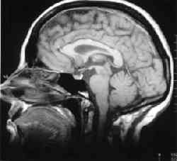
MRI stands for Magnetic Resonance Imaging. This type of scan uses magnetism to build up a picture of the inside of the body. Below is an example of an MRI scan of the head.

MRI is completely painless, but the scanner is very noisy. The MRI scanner creates cross section pictures of the body. It can show up soft tissues very clearly and a single scan can produce many pictures from angles all round the body. They can be affected by movement so they aren’t used very often for some tumours because coughing, swallowing or breathing will make the scan less clear.
MRI can be used on most areas of the body. For some parts of the body and for some types of tissues, it can produce clearer results than a CT scan. For other situations, the CT scan is better. Your own doctor will know which is the best type of scan for you.
MRI is particularly good for some types of brain tumour, primary bone tumours, soft tissue sarcomas and for tumours affecting the spinal cord. In some situations, your doctor may suggest MRI if a CT scan hasn’t been able to give all the information they need. In some early cancers, such as cervix or bladder cancer, MRI is better than CT at showing how deeply the tumour has grown into body tissues. It can be particularly useful for showing whether tissue left behind after treatment is tumour or not.
As well as being used to find or stage tumours, MRI can be used to measure blood flow. You may have a type of special dye by injection before the scan to help make the pictures clearer.
READ MORE: http://cancerhelp.cancerresearchuk.org/about-cancer/tests/mri-scan
|
READ MORE: http://www.breastthermography.com/


 The use of Digital Infrared Imaging is based on the principle that metabolic activity and vascular circulation in both pre-cancerous tissue and the area surrounding a developing breast cancer is almost always higher than in normal breast tissue. In an ever-increasing need for nutrients, cancerous tumors increase circulation to their cells by holding open existing blood vessels, opening dormant vessels, and creating new ones (
 Current methods used to detect suspicious signs of breast cancer depend primarily on the combination of both physical examination and mammography. While this approach has become the mainstay of early breast cancer detection, more is needed. Since the absolute prevention of breast cancer has not become a reality as of yet, efforts must be directed at detecting breast cancer at its earliest stage. As such, the addition of Digital Infrared Imaging (Breast Thermography) to the frontline of early breast cancer detection brings a great deal of good news for women.